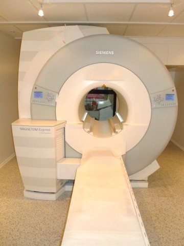
Magnetic Resonace Tomogrphy (MRI)
The MRI is a technically highly complex, yet absolutely harmless and very sensitive way to examine the human body. The method is based on the combined use of a strong magnetic field together with radiowaves. In medical use it hasn´t any side-effects and it is completely free of any radiation or radioactivity.
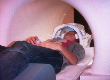
During the examination the part of the body which has to be examined has to be positioned into the middle of a ring with a diameter of about 70 cm and a length of about 100 cm. This ring is open all the time and is very well air-conditioned.
While the examination is in progress a loud and repetitive noise is heard, this is why you are offered ear-protection before the start of examination.
When you hear the noise it means, that the measurement is made at that time. Each measurement will take about 3-4 minutes. In between these measurements there will be pauses of a few seconds up to some minutes, in this time the gathered information is stored. The whole examination will take right from the start up to end about 20 to 40 minutes.
From the time you enter the practice up to the end of the explanation of your diagnosis to you by the doctor usually one hour has passed.
For some examinations it is necessary to inject a contrast-media (about 20 ml Gadolineum) in an vein of the arm. Side-effects of this contrast-media are very rare. The contast-media spreads in the whole body with the blood-circulation within a few seconds. For fresh injuries a contrast-media is usually not needed. But for the examination of the brain, the abdomen or in case of chronic pain or after operations it is usually very helpful to make the diagnosis more precise.
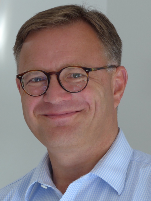
Dr. med. Walter Riffeser
1959 - born and grown up in Munich
1979 - 1980 Military service in 1st Mountain-Division
1980 - 1985 Study at Ludwig-Maximilians-Universität
Munich
1986 - Surgery at University of Cambridge, England
- Medicine at Medizinische Poliklinik, Munich
- Orthopedic surgery at Staatliche Orthopädische Klinik
Munich
1986 - Thesis on hip-dislocation in children with cerbral palsy
(Prof. Dr. Stotz)
1986 - 1987 Orthopedic Surgery (Prof. Dr. H. Täger) and
Pathology at Klinikum Großhadern (Prof. Dr. Eder)
1987 - 1989 General surgery at Chirurgische Klinik Dr. Rinecker
1990 - 1995 Diagnostic Radiology at the University-Hospitals of
Rechts der Isar and Großhadern, both in Munich
1996 - Final exam for Diagnostic Radiology
1997 - Partner of a radiological practice in Munich
2007 - Foundation of Practice Maximilianstraße